

November 15, 2021
by Gabriele Maycher, CEO, GEM Dental Experts Inc. BSc, PID, dip DH, RDH
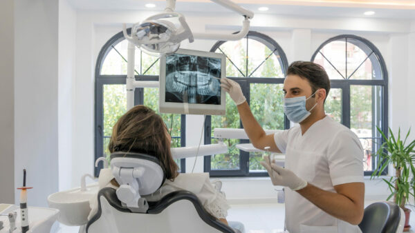
- It’s important to keep radiation exposure as low as possible while still gathering the needed radiographic evidence. So how low is too low?The first thing that typically comes to mind when discussing imaging and the risk of exposure to ionizing radiation is “ALARA,” an acronym for “As Low as Reasonably Achievable.” This means we need to make every reasonable effort to maintain exposures to ionizing radiation as far below the dose limits as practical for a diagnosis and evaluation.
I would suggest that if you can’t make an accurate dental hygiene diagnosis (DHD), the number of radiographs you’re taking is probably too low. Radiographic assessment forms a critical component of clinical assessment, as periodontal assessments alone simply don’t tell the whole story. And without an accurate diagnosis, there can be no logical treatment plan, effective implementation, and evaluations. As you can see, the process of care simply falls apart right from get-go.
EXPOSING A CHRONIC PROBLEM
In my experience, radiographic evidence is lacking and/or inconsistent. When conducting chart audits, I rarely find adequate radiographs to accurately screen for the potential periodontal conditions that may exist in any given patient. The other problem: The type of radiographs being exposed (i.e., horizontal bitewings vs. vertical bitewings) and the diagnostic quality of the radiographs (i.e., overlap and the inability to see the lamina dura or the alveolar crestal bone height on either the maxilla, the mandible, or both) makes me wonder if the radiographs are being evaluated for a DHD at all! This hunch is typically confirmed by an absence of documented radiographic interpretation.Although we have the American Dental Association (ADA) guidelines www.ada.org as a reference for prescribing radiographs, we still need to use clinical judgement when determining which radiographs are required. This clinical judgement is subject to understanding the current evidence-based literature.
To fully ensure we are screening for all possible periodontal diseases and conditions, we need to understand the characteristics of each of these diseases. So, let’s breakdown the categories in the 2018 AAP Periodontal Classification (Table 1: 2018 AAP Periodontal Classification) and see if we are, in fact, taking adequate radiographs.
Table 1: 2018 AAP Periodontal Classification

Category: Periodontal health, gingival disease and conditions
The first information your four diagnostic vertical BWs will reveal is what type of periodontium exists. This will be paramount in determining your DHD (health, gingival disease, periodontitis) and how you will move forward with the process of care. We are looking to identify one of the following:Intact periodontium. Radiographic features of a normal, anatomically intact periodontium would include an intact lamina dura (both laterally and at the alveolar crest), no evidence of bone loss in furcation areas, and approximately 2mm distance on average (can vary between 1.5mm–3mm) from the most coronal portion of the alveolar bone crest (AC) to the cementoenamel junction (CEJ).1
Of course, you can’t truly know if you have an intact periodontium with just a set of BWs. What about the anteriors? Exposing anterior (maxilla and mandibular) periapicals (PAs) to screen for what was formally known as aggressive or juvenile periodontitis may be prudent. This condition has now been collapsed into the category of periodontitis and incorporated into the Staging and Grading process.
The American Academy of Periodontology (AAP) agreed there was no evidence to suggest a specific pathophysiology that enabled differentiation of aggressive and chronic.2 They do, however, recognize that the clinical presentation differs based on age of the patient, number of lesions, severity, and location within the dental arch.2 Hence, they have incorporated these observations within the Staging and Grading system by adding a molar/incisor pattern in addition to localized/generalized descriptors for extent/distribution of periodontitis. As well, the grading classification system has accommodated periodontitis with higher rates of progression for patients presenting with more severe destruction at an earlier age2 (aggressive/juvenile periodontitis) with a Grade C score (i.e., destruction exceeds expectation of biofilms). A minimum of two anterior PAs may screen for this.
Reduced periodontium due to non-periodontitis or acquired and developmental conditions like recession due to cervical restorations, or alveolar bone loss due to ortho forces, open contacts, extractions, crown lengthening, etc. In conjunction with the patient’s medical dental history, as well as periodontal and hard-tissue assessments, the clinician should be able to determine if a reduced periodontium due to non-periodontitis exists. Again, if the reduced periodontium is due to non-periodontitis causes, an additional set of anterior PAs may be warranted to screen for the potential of Grade C periodontitis or other causative factors reducing the periodontium in the anterior region.
Reduced periodontium due to periodontitis. This is defined as alveolar bone loss due to biofilms initiating an inflammatory response. If the reduced periodontium is due to periodontitis, the AAP recommends a FMS as part of the initial case overview to assess disease. See Table 2: Three Steps to Staging and Grading.
Table 2: Three Steps to Staging and Grading

For patients who have a reduced periodontium due to periodontitis and localized or generalized areas of breakdown due to non-periodontitis causes a FMS is still required.
Category: Periodontitis
I rarely see a FMS for a patient with periodontitis, but this step is vital for the following reasons:- To establish a baseline of the current state of periodontal breakdown. Patients with periodontitis have periods of disease activity and inactivity, or remission. Tissue destruction will progress at different rates throughout the mouth, and destruction may not occur in all parts of the mouth at the same time. Some sites may remain unchanged for long periods while others may progress more rapidly as seen in random burst patterns or multiple burst patterns.3,4 A stable periodontitis patient remains at higher risk for recurrent disease compared with a gingivitis patient or a healthy patient. Therefore, ongoing, individual monitoring and risk assessment as part of optimal patient management is required.5
- To Stage and Grade a patient. When radiographic bone loss (RBL) vs. clinical attachment loss (CAL) is used as a stage determinant, a FMS as an overview of the dentition is required to determine the site of greatest loss interdentally (attributable to periodontitis only). RBL is measured in percentage of root length, so bitewings alone don’t provide enough of an overview to make this calculation.
A FMS also assists in determining Grade. You can determine grade by using either direct or indirect evidence. Direct evidence is based on the longitudinal observation available from older diagnostic quality radiographs2 (i.e., previous PAs, FMS comparison). Indirect evidence is based on the assessment of RBL at the worst affected tooth in the dentition as a function of age (RBL measured in percentage of root length divided by age of the subject).2 Again, you will need a FMS to make this calculation.
We know that periodontitis grade may be modified by the presence of such risk factors as smoking and diabetes, but if none exist, you will need to rely on direct or indirect evidence.
Approximately 47% of patients have periodontitis. In adults 65 and older, prevalence rates increase to 70%.7 So, chances are 50% or more of your patients may need a FMS to establish an accurate DHD.
Category: Other conditions affecting the periodontium
Additional PAs may be required in this category to identify causes that may contribute to the breakdown of the periodontium. They include periodontal abscesses and endodontic-periodontal lesions, tooth- and prosthesis-related factors (bulbous crowns, overhangs, supracrestal impingement, etc.), mucogingival deformities, and conditions like inadequate attached gingiva to name a few.Category: Peri-Implant Disease and Conditions
Your dentist will establish a radiographic baseline at the time of implant placement, but monitoring the implant’s health may require additional PAs at the first signs of inflammation to rule out peri-implantitis. The onset of peri-implantitis may occur early during follow-up, and progress in a non-linear and accelerated pattern.6PANORAMIC RADIOGRAPH
As per AAP, (perio.org) a pano-ramic (PAN) radiograph is useful for screening bone loss patterns in general, pathologies and anomalies, cysts, and abscesses, to monitor growth and development of the jaw and wisdom teeth. As well, it provides a general view of the oral structure. The PAN completes the comprehensive screening of periodontal disease and much more.Visit the ADA website (ada.org) for panoramic guidelines.
SETTING THE BAR HIGH
So far, I have only discussed the lack of radiographs required for an accurate DHD, but what about the radiographs required to support the phases of implementation and evaluation in nonsurgical periodontal therapy? Again, I would debate if adequate radiographs are exposed in these phases to assist in achieving endpoint, but we’ll save this discussion for another article.Lastly, I know the argument can always be made that patients don’t want even the minimum number of radiographs recommended. My response? If you don’t create value, of course they don’t. By giving evidence-based rationale for why the radiographs are required, explaining the importance of screening for all periodontal conditions that may exist, and educating your patients on the oral and systemic link, you will create value and acceptance. But perhaps even more important, you as the clinician need to value the vital role radiographs play in every step of the process of care and leverage them to achieve superior patient outcomes!
Are radiographs in your Scope of Practice?
As clinical hygienists, we have all been educated in diagnosing/identifying periodontal disease, identifying normal from abnormal findings, and interpreting radiographs for the purpose of a dental hygiene diagnosis. However, only six provinces and territories allow hygienists to prescribe radiographs for a DHD, so if radiographic imaging is not in your scope of practice, the dentist is responsible for prescribing the radiographs needed to make a diagnosis and verifying your findings. For more information, visit https://files.cdha.ca/profession/Regulatory_Authority_chart_0620.pdf
References
- Lang NP, Bartold PM. Periodontal health. J Periodontol.
2018;89(Suppl 1): S9–S16. https://doi.org/10.1002/JPER.16-0517 - Tonetti MS, Greenwell H, Kornman KS. Staging and grading of periodontitis: Framework and proposal of a new classification and case definition. J Periodontol. 2018;89(Suppl 1):S159– S172. https://doi.org/10.1002/JPER.18-0006
- Lindhe J, Okamoto H, Yoneyama T, Haffajee A. Socransky SS. Longitudinal changes in periodontal disease in untreated subjects. J Clin Periodontol. 1989;16 (10): 662-670.
- Kakuta E, Nomura Y, Morozumi T, et al. Assessing the progression of chronic periodontitis using subgingival pathogen levels: a 24-month prospective multicentre cohort study. BMC Oral Health. 201717(1):46.
- Chapple ILC, Mealey BL, et al. Periodontal health and gingival diseases and conditions on an intact and a reduced periodontium: Consensus report of workgroup 1 of the 2017 World Workshop on the Classification of Periodontal and Peri-Implant Diseases and Conditions. J Periodontol. 2018;89(Suppl 1):S74–S84. https://doi.org/10.1002/JPER.17-0719
- Renvert S, Persson GR, Pirih FQ, Camargo PM. Peri-implant health, peri-implant mucositis, and peri-implantitis: Case definitions and diagnostic considerations. J Periodontol. 2018;89(Suppl 1):S304–S312. https://doi.org/10.1002/JPER.17-0588.
- P.I. Eke, B.A. Dye, L. Wei, G.O. Thornton-Evans, and R.J. Genco. Prevalence of Periodontitis in Adults in the United States: 2009 and 2010. J DENT RES 0022034512457373, first published on August 30, 2012 as doi:10.1177/0022034512457373


Leave a Reply Cancel reply
You must be logged in to post a comment.